José María Carazo
Group Leader
Carlos Oscar Sorzano
Group Leader
Research summary
We work in the field of cryo Electron Microscopy, constantly exploring ways to maximize the structural information that can be extracted from the images by novel image processing approaches. We develop new methods and new software, applying it to the analysis of purified macromolecules (the so called "Single Particle Analysis") as part of our Instruct facility access role. Currently we are diving more and more into cryo EM Tomograpgy, focusing into membrane complexes in their native environment.
Publications
Melero R, Sorzano COS, Foster B, Vilas JL, Martínez M, et al. Continuous flexibility analysis of SARS-CoV-2 spike prefusion structures. IUCrJ 2020; 7 (6): 1059-1069.
Carter SD, Hampton CM, Langlois R, Melero R, Farino ZJ, Calderon MJ, Li W, Wallace CT, Tran NH, Grassucci RA, Siegmund SE, Pemberton J, Morgenstern TJ, Eisenman L, Aguilar JI, Greenberg NL, Levy ES, Yi E, Mitchell WG, Rice WJ, Wigge C, Pilli J, George EW, Aslanoglou D, Courel M, Freyberg RJ, Javitch JA, Wills ZP, Area-Gomez E, Shiva S, Bartolini F, Volchuk A, Murray SA, Aridor M, Fish KN, Walter P, Balla T, Fass D, Wolf SG, Watkins SC, Carazo JM, Jensen GJ, Frank J, Freyberg Z. Ribosome-associated vesicles: A dynamic subcompartment of the endoplasmic reticulum in secretory cells Science Advances. 2020 6, eaay9572
Coloma R, Arranz R, de la Rosa-Trevin JM, Sorzano COS, Munier S, Carlero D, Naffakh N, Ortín J, Martin-Benito J.The processive helical track: A structural model for influenza virus transcription and replication. Nat Microbiology 2020; 5: 727-734
Vilas JL, Tagare HD, Vargas J, Carazo JM,Sorzano COS. Measuring local-directional resolution and local anisotropy in cryoEM maps. . Nature Communications 2020; 6: 21626
Ramírez-Aportela E, Mota J,Conesa P, Carazo JM, Sorzano COS. DeepRes: A New Deep Learning and aspect-based Local Resolution Method for Electron Microscopy Maps. IUCR J 2019; 11: 55
Segura J, Sanchez-Garcia R, Sorzano COS, Carazo JM. 3DBIONOTES v3.0: Crossing molecular and structural biology data with genomic variations. Bioinformatics 2019; 35: 3512-3513
The 2013-2014 biennium was particularly important for the field of cryogenic electron microscopy (cryo EM). Thanks to key technological developments in instruments and software, it became obvious in 2013 that cryo EM had indeed matured as a technique, such that we can now systematically produce quasi-atomic structural information for a wide range of very challenging macromolecular complexes. The need to re accommodate all image processing workflows to address the new challenges in the field consequently became the target of the group. This goal fitted very well with our focus on methods and software development in our role as the image processing infrastructure providers for Instruct, the structural biology project of the European Strategic Forum for Research Infrastructures.
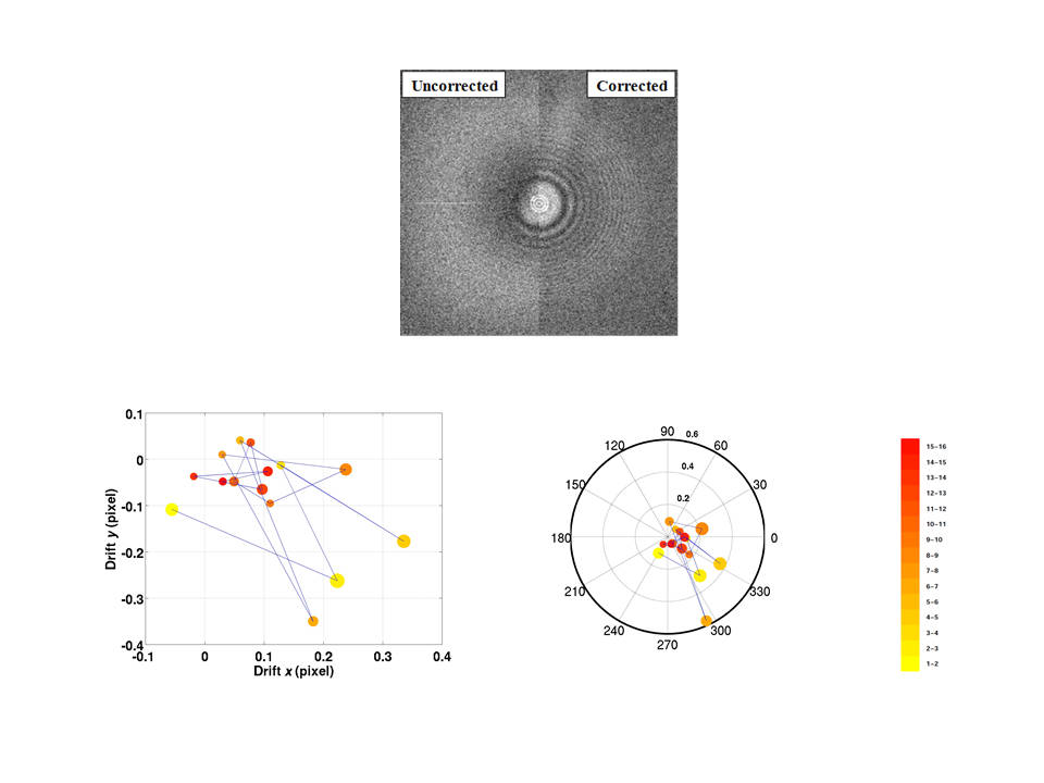
We have explored new areas of cryo EM image processing within our traditional software suite XMIPP (downloaded in recent years by thousands of researchers from all over the world). We addressed a number of topics central to enhanced microscope performance and optimal image processing, such as accurate, parameter-free optical characterization, automatic analysis of micrographs, or image-ranking procedures, all critical to render cryo EM a high throughput technique for structural biology. With regard to software, we introduced the beta version of our key development, Scipion, a graphic image-processing workflow integrator that assures full tracking of all analytic results, while offering an environment that integrates most of the best known software packages in the field.
With respect to X-microscopy, using the X-ray microscope at the Spanish synchrotron ALBA, we produced the first detailed optical characterization with this type of instrument, while advancing in automation and image processing capabilities.
Software developed by the group and available for the Scientific Community
![]()
An image processing framework for 3D Electron Microscopy.

Web application designed to automatically annotate biochemical and biomedical information onto structural models
 A suite of image processing programs for single-particle 3D Electron Microscopy & more