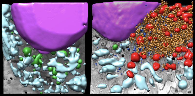
Pablo Gastaminza, CNB-CSIC researcher and main author of the work, explains the alterations they found: "when comparing an uninfected cell with an infected one, we can see that the virus multiplication machinery forms vesicles and tubules as well as remarkable signs of stress on cellular organelles such as mitochondria and the endoplasmic reticulum."
The study is part of the collaboration established within the European CoCID (Compact Cell Imaging Device) consortium. It combines the use of molecular biology, virology and three types of microscopy techniques. One of them is the so-called soft X-ray cryo-tomography (Cryo-SXT), a technology available only in four places all over the world, including the MISTRAL beamline at the ALBA Synchrotron. This technique allows "to generate three-dimensional maps of the ultrastructure of complete cells, reconstructing their total volume and providing extra information to other techniques like electron microscopy," according to Eva Pereiro, head of the MISTRAL beamline at ALBA.
Ana Joaquina Pérez-Berná and Victoria Castro, first authors of the study, from the ALBA Synchrotron and the CNB-CSIC respectively, assure that "obtaining these images is of great help to better understand how the virus works, since they show how it takes advantage of parts of the cell's cytoskeleton (such as vimentin and centrioles) to use them as a kind of ‘scaffold’ and build their virus replication factory there." Knowing these processes enable to develop and better comprehend therapies to fight the virus.
Scientific reference
Victoria Castro, Ana Joaquina Pérez-Berna, Gema Calvo, Eva Pereiro, and Pablo Gastaminza. Three-Dimensional Remodeling of SARS-2 CoV2-Infected Cells Revealed by Cryogenic 3 Soft X‑ray Tomography. ACS Nano. DOI: 10.1021/acsnano.3c07265
CNB, CSIC Comunicación, ALBA Comunicación