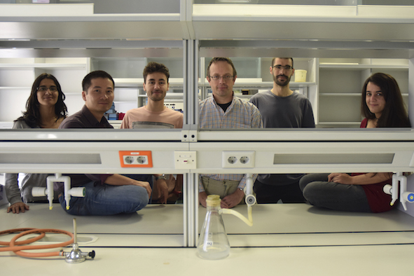
members (left-to-right)
Marta Sanz Gaitero - doctoral student (RISAM fellowship)
Thanh H. Nguyen - postdoc
Mateo Seoane Blanco - doctoral student (FPI fellowship)
Mark J van Raaij - cientifico titular
Antonio Pichel Beleiro - doctoral student (RISAM fellowship)
Mara Laguna - technician
Some viruses and bacteriophages attach to their host cell via proteins integral to their capsids, for example poliovirus, coxsackievirus and rhinovirus ('common cold virus'). Other viruses bind to their host cell receptors via specialized spike proteins (for example HIV, the AIDS-virus), or via specialized fiber proteins, like adenovirus, reovirus and many bacteriophages.
Virus fibers have the same basic architecture: they are trimeric and contain an N-terminal virus or bacteriophage attachment domain, a long, thin, but stable shaft domain and a more globular C-terminal cell attachment domain. Their detailed folds are very diverse and often reveal novel features. These trimeric, fibrous proteins are very stable to denaturation by temperature or detergents. Our goal is to determine the structures of these proteins and thus to make an extensive inventory of stable trimeric folds present in nature. We also want to explain how they bind their receptors and therefore aim to crystallize them with receptor analogues.
news
february 2018: mini-review about contractile tail systems published in Molecular Microbiology
january 2018: Thanh left to go back to the Vietnamese Academy of Science and Technology Institute of Biotechnology in Hanoi - we wish him a lot of success!
december 2017: review on Bacteriophage T4 Long Tail Fiber Domains online
october 2017: paper including snake adenovirus LH3 protein structure published in Structure
september 2017: CASP12 Target Highlights paper online
june 2017: paper on phage T4 gp34 structure published in Viruses (on the cover)
april 2017: Mark appointed Section Editor of Acta Crystallographica F
structure gallery
bacteriophage fibers
adenovirus proteins
bacteriophage endolysins
bacterial dehydroquinase, shikimate kinase
collaborators
contact
group webpage @ CNB-CSIC
positions

Section Editor of Acta Crystallographica F, Structural Biology Communications
ResearchGate page Mark van Raaij