Roberto Vázquez and colleagues at the CIB-CSIC found that this protein possesses enzymatic activity of the muramidase variety, capable of degrading the glycan chains of the bacterial cell wall peptidoglycan. They also characterized the specific amino acids involved in this catalytic activity.
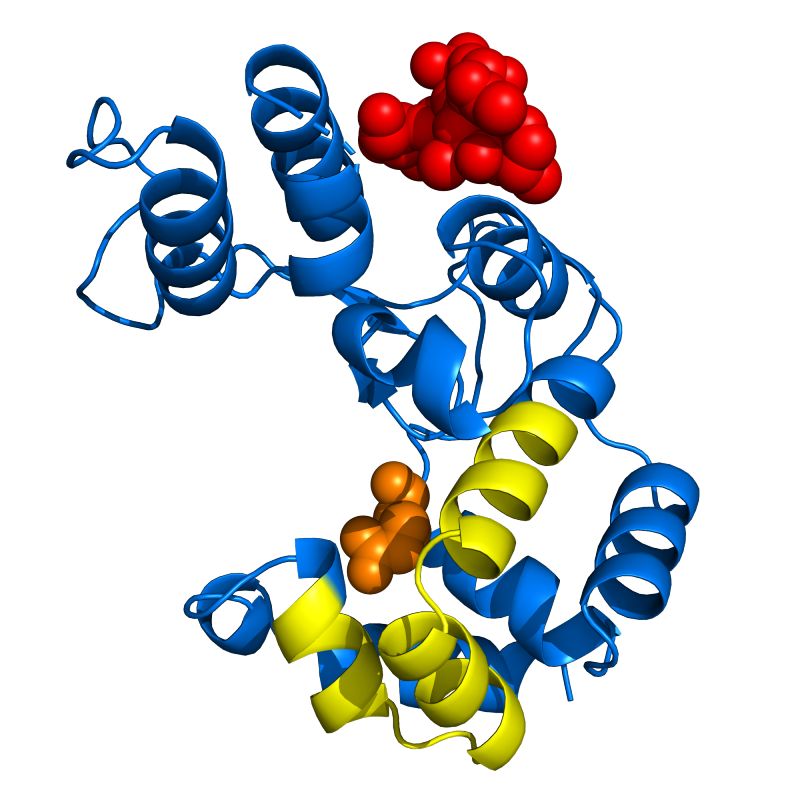
Mateo Seoane-Blanco at the CNB-CSIC crystallized the protein and solved its structure using the X-ray crystallography beamline BM13-XALOC of the ALBA synchrotron in Barcelona. The novelty in the Pae87 structure is that even though its general structure is similar to other endolysins, it lacks a cell-wall binding domain. According to van Raaij, “a simple but elegant experiment performed was to incubate the enzyme with purified peptidoglycan and then also crystallizing the resultant mixture, which also led to the structure of endolysin in complex with a peptidoglycan fragment”. This complex structure revealed that Pae87 has a separate substrate-binding region within the catalytic domain itself. This substrate-binding region is conserved among related endolysins lacking an additional cell wall-binding domain, but not among those containing such a module, and thus may serve a compensatory evolutionary function.
The CIB group also found that endolysin Pae87 possesses a non-enzymatic activity that acts on the membranes of Gram-negative bacteria, which most probably resides in a peptide near the end of the lysin sequence called P87 (in yellow in the accompanying figure). Alone, this P87 peptide exhibits potent antibacterial activity.
These results open the way for future efforts to design and obtain antimicrobials based on phage endolysins, not only by modifying the whole enzymes, but also by deriving small antimicrobial peptides from their structure.
Scientific reference
Monomodular Pseudomonas aeruginosa phage JG004 lysozyme (Pae87) contains a bacterial surface-active antimicrobial peptide-like region and a possible substrate-binding subdomain. Roberto Vázquez, Mateo Seoane-Blanco, Virginia Rivero-Buceta, Susana Ruiz, Mark J. van Raaij, and Pedro García (2022), Acta Crystallographica section D, volume 78, pages 435-454, doi.org/10.1107/S2059798322000936