A study published recently in the journal PLoS Pathogens shows that throughout evolution, the double-stranded RNA viruses of fungi have acquired new enzyme activities on their outer surface. The publication is the result of an international collaboration led by scientists at the Centro Nacional de Biotecnología of the CSIC (CNB-CSIC).
Most viruses that infect fungi are transmitted directly from cell to cell, with no exposure to the exterior. In plant, animal and bacterial viruses, the primary function of the virus capsid is to protect the genetic material and identify its hosts, but in fungal viruses these functions are less important. For this reason, the external capsid has different structural and functional characteristics.
"Evolution has allowed modifications in very specific domains of the proteins that form the capsid. This led to new functions that allow the viruses to maintain a closely regulated symbiotic relationship with the cells they infect," explains José R. Castón, lead author of the study and CNB-CSIC researcher.
To reach this conclusion, the authors studied the atomic structure of the proteins that form the viral capsid and identified two domains with enzyme activity. "These active domains seem, in one case, to be able to bind proteins of the cell cytoskeleton, and in the other, to interact with and cleave peptides," says Castón.
To determine the structure of these proteins at atomic resolution, they used the electron cryomicroscopy technique – which earned the Nobel Prize in Chemistry in 2017.
"The potential of fungal viruses as molecular nanomachines has not yet been explored, but the peculiarities of their capsid could make them interesting tools for biotechnological applications," Castón concludes.
- Mata CP, Luque D, Gómez-Blanco J, Rodríguez JM, González JM, Suzuki N, Ghabrial SA, Carrascosa JL, Trus BL, Castón JR. Acquisition of functions on the outer capsid surface during evolution of double-stranded RNA fungal viruses PLoS Pathog. 2017 Dec 8;13(12):e1006755.

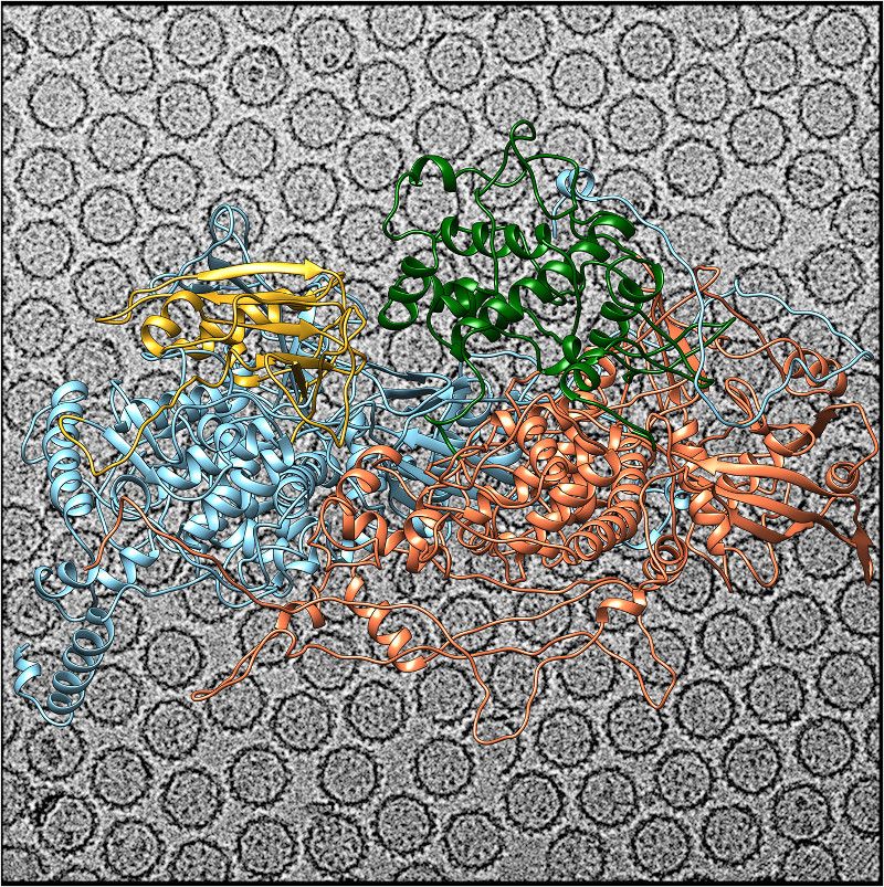
La cápsida de Rosellinia necatrix quadrivirus 1 (RnQV1) es un icosaedro T=1 formado por heterodímeros de P2 (azul) y P4 (naranja). Inserciones de dominios en P2 y P4 proporcionan funciones adiccionales en la superficie externa de la cápsida. La inserción en P2 tiene un plegamiento similar a proteínas relacionadas con el metabolismo del citoesqueleto celular (amarillo); la inserción en P4 sugiere una actividad proteasa implicada en el procesamiento de P2 (verde). El fondo muestra una imagen de criomicroscopía electrónca de RnQV1. / José R. Castón CNB-CSIC